Back Of Skull And Neck Anatomy : Human Body Skull Anatomy External Occipital Protuberance Human Back Of Skull Face Human Png Pngegg / Anterior (ossified within months) leads to stifness of the neck due to fibrosis and shortening of the sternocleidomastoid.
Back Of Skull And Neck Anatomy : Human Body Skull Anatomy External Occipital Protuberance Human Back Of Skull Face Human Png Pngegg / Anterior (ossified within months) leads to stifness of the neck due to fibrosis and shortening of the sternocleidomastoid.. 3 skull continued **fontanels in the skull are the unossified remnants of the membranes in newborns. In fact, there are twenty three in total, some of which are paired Bones of the neck picture. Human a skull consists of the frontal, temporal, parietal and occipital bones. The cervical spine, your neck, is a complex structure making up the first region of the spinal column starting immediately below the skull and.
Passing back and slightly upwards from this foramen is where the external oblique line, which becomes continuous with the. All the bones of skull, joined together by sutures… anatomy ▶ head and neck ▶ bones and cartilages ▶ skull. In radiology, the 'head and neck' refers to all the anatomical structures in this region excluding the central nervous system, that is, the brain and spinal co. It joins the parietal bones at the. .back of the neck, and laterally it is prolonged into the temporal region, where it is looser in texture.
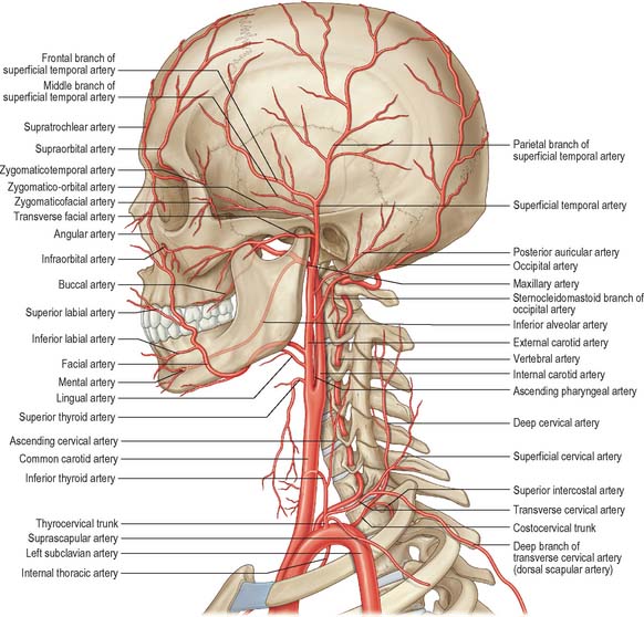
Nerves And Arteries Of Head And Neck Anatomy Branches Kenhub from thumbor.kenhub.com Instant anatomy is a specialised web site for you to learn all about human anatomy of the body with diagrams, podcasts and revision questions. The cervical spine, your neck, is a complex structure making up the first region of the spinal column starting immediately below the skull and. .back of the neck, and laterally it is prolonged into the temporal region, where it is looser in texture. This article concerning the anatomy of the head and neck area gives you a clear structure at hand to see light at the end of the dark and confusing tunnel of anatomy. In radiology, the 'head and neck' refers to all the anatomical structures in this region excluding the central nervous system, that is, the brain and spinal co. The occipital bone forms the back and base of the cranium ( fig. The skull or known as the cranium in the medical world is a bone structure of the head. Muscle head anatomy vocal organ diagram female neck anatomy neck wireframe head neck human anatomy head artery anatomy face pharynx vector neck degree head anatomy 3d.
The splenius muscles originate at the midline and run laterally and superiorly to their insertions.
Instant anatomy is a specialised web site for you to learn all about human anatomy of the body with diagrams, podcasts and revision questions. How many moveable vertebrae are in the… what are the main purpose of transverse… The trapezius originates from the skull and spine of the upper back and neck. I was operated 3 years ago from my neck and got 3 bones removed it is the most painful feeling a human will ever experience i now have titanium plates. Anatomy of the head and neck. The skull is a bony structure that supports the face and forms a protective cavity for the brain. Top head neck anatomy flashcards ranked by quality. Cranial cavity , cranial sutures. The skull is composed of 28 separate bones, most of them paired (ch. The erector spinae are a group of many muscles that attach along the back of the spine. This article describes the anatomy of the head and neck of the human body, including the brain, bones, muscles, blood vessels, nerves, glands, nose, mouth, teeth, tongue, and throat. This is the uppermost of the cervical vertebrae. It attaches to the clavicle and scapula.
I thought i'd use this channel to share some anatomy thoughts and include some of the other stuff too. Foramina of the skull and the structures that pass through. The head rests on the top part of the vertebral column, with the skull joining at c1. Navigate through the head and neck by the by type of body part you are looking for. The foramen magnum, housing the brainstem, is also a part of the occipital bone.
Triangles Of The Neck Anatomy Borders And Contents Kenhub from i.vimeocdn.com It contains an external occipital protuberance that can be felt on the back of your head. (anatomy of the head and neck): The dentist is most concerned on the maxillary bones. Bones of the skull and more skull anatomy. It attaches to the clavicle and scapula. There is a superficial layer of fascia in the neck, and there are 3 deep layers of fascia. Human a skull consists of the frontal, temporal, parietal and occipital bones. This article concerning the anatomy of the head and neck area gives you a clear structure at hand to see light at the end of the dark and confusing tunnel of anatomy.
They don't move and united into a single unit.
They are divided into three layers. It contains an external occipital protuberance that can be felt on the back of your head. From the sides and the back of the neck, the splenius capitis inserts onto the head region, and the splenius. Human a skull consists of the frontal, temporal, parietal and occipital bones. Muscle head anatomy vocal organ diagram female neck anatomy neck wireframe head neck human anatomy head artery anatomy face pharynx vector neck degree head anatomy 3d. Excluding ear ossicles, it is made of 22 bones. The skull is the bony skeleton of the head. Want to learn more about it? Tendons are connective tissue that attach muscle to bone, whereas ligaments attach bones to other bones. Neck muscles work together with tendons and ligaments to support and move the neck and head. Anatomy, head neck anatomy, medical & nursing. Injury during delivery may also result in torticollis. (anatomy of the head and neck):
It contains an external occipital protuberance that can be felt on the back of your head. Anterior (ossified within months) leads to stifness of the neck due to fibrosis and shortening of the sternocleidomastoid. How many moveable vertebrae are in the… what are the main purpose of transverse… .back of the neck, and laterally it is prolonged into the temporal region, where it is looser in texture. Navigate through the head and neck by the by type of body part you are looking for.
Head And Neck Overview And Surface Anatomy Basicmedical Key from basicmedicalkey.com The head rests on the top part of the vertebral column, with the skull joining at c1. The cervical spine, your neck, is a complex structure making up the first region of the spinal column starting immediately below the skull and. The occipital bone forms the back and base of the cranium ( fig. The muscles of the back and neck are responsible for maintaining posture and facilitating movement of the head and neck. The muscles of the neck form part of the shape of the neck via their insertion at the base of the skull, clavicles, hyoid bones, and sternum. I teach human anatomy and do a bunch of other things in my life. Injury during delivery may also result in torticollis. Third, fourth and fifth cervical nerves innervate the scalp and the skin over the back of the neck, and.
.and neck (such as the complex infratemporal fossa area) are not readily familiar to even the most experienced of surgeons, who may have to refer to anatomy texts and a dried skull before operating in this region.
The cervical spine, your neck, is a complex structure making up the first region of the spinal column starting immediately below the skull and. There is a superficial layer of fascia in the neck, and there are 3 deep layers of fascia. The occipital bone forms the back and base of the cranium ( fig. It contains an external occipital protuberance that can be felt on the back of your head. The foramen magnum, housing the brainstem, is also a part of the occipital bone. Head and neck anatomy is important when considering pathology affecting the same area. The major sutures are the coronal suture, sagittal suture, lambdoid suture and squamosal sutures. They are divided into three layers. How many moveable vertebrae are in the… what are the main purpose of transverse… The splenius muscles originate at the midline and run laterally and superiorly to their insertions. The skull is a bony structure that supports the face and forms a protective cavity for the brain. Muscle head anatomy vocal organ diagram female neck anatomy neck wireframe head neck human anatomy head artery anatomy face pharynx vector neck degree head anatomy 3d. Foramina of the skull and the structures that pass through.
It contains an external occipital protuberance that can be felt on the back of your head back of skull anatomy. The muscles of the back and neck are responsible for maintaining posture and facilitating movement of the head and neck.


0 Comments